Wednesday, July 29, 2009

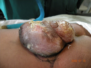
Cirsoid aneurysm of scalp


This young guy presented with pulsatile swelling in the scalp
O/E continuous machinery murmur could be heard over the swelling
Diagnosis Cirsoid aneurysm of scalp
Exploration of the pulsatile mass with visualisation of the feeding artery done and the ligation of the feeding vessel with excision of the dilated vessels done

Fourth Ventricle tumor


Cerebelar tonsil retracted
Brain stem visualised nicely
PICA is seen in the operating field


midline incision from inion to level of C2
Occiput and 1st dorsal arch and 2nd cervical spine visualised


This is a case of 16 yrs old female presenting with headache , dizziness for 1 yr .
Case was operated and a firm mass was visualised in fourth ventricle reaching up t0 mid brain .
Suboccipital craniectomy was done and 1st dorsal arch also removed Y shaped incision over dura . Cerebellar tonsil retracted and vermis exposed and retracted tumor mass was visualised and total resection of tumor from ventricular bed along with its extension achieved .

Monday, July 27, 2009

Cerebello pontine angle Tumor


Cerebellopontine angle is the region where petrosal surface of cerebellum wraps around the pons and upper medulla with the middle cerebellar peduncle being its floor .
CPA is formed by superior and inferior limbs of cerebellopontine fissure with the apex oriented laterally .
It is the site for 8 to 10 % of intracranial tumor.
Most common tumor at this area are Vestibular Schwannoma (80 to 90 %) , Meningioma and Epidermoid cyst.
Anatomy of CPA can be conceptualized by breaking down various neurovasculature into 3 complexes .
Upper complex consists of V CN , superior cerebellar artery and petrosal vein.
Middle coplex consists of VII and VIII CN complex and Anterior Inferior Cerebellar Artery (AICA) and its surgically important 3 branches internal auditary artery ( labyrinthine artery ) , the recurrent perforating artery and subarcuate artery .
Lower complex consists of IX X XI CN complex and Posterior Infereior cerebellar artery. ( PICA)

Friday, July 24, 2009

Brain Abscess Or Tumor


Sometimes its so confusing to distinguish brain abscess and brain tumor on scans . Ring enchancing lesion on scan.
MRI spectroscopy helps to distinguish between two
What is Magnetic Resonance Spectroscopy?

Simply put, MRS is a quantitative method of display of non-aqueous proton signals that correspond to certain biological molecules in the brain.
Magnetic resonance imaging (MRI) uses water protons to create images. Very sophisticated and detailed images of the brain are produced based on the fact that different tissues within the brain have different water content. The hydrogen nuclei (protons) that are not in water (non-aqueous protons) are distributed throughout the hundreds of biologically significant molecules of the living brain. These molecular radio-signals are not visible on standard MR images because they are overwhelmed by the high signal derived from the aqueous protons. The non-aqueous molecules each has a unique radio-frequency specific to the chemical itself and distinct from the frequency of water protons. The amplitude or strength of these radio-frequencies is dependent on their concentration in the volume of interest, each at their discrete radio-frequence position. This allows a graph to be displayed that shows the relative peaks of amplitude for each of several biological molecules. These peaks are more readily detected and displayed if the signal from aqueous protons is suppressed.
What is MR Spectrum?
The "spectrum" is a graph of the relative concentrations of these molecules based on their discrete radio-frequency signal, which is shifted away (chemical shift) from the water signal the MRI uses. A high peak means that the molecule at that RF location exists in greater concentration than an adjacent low peak. To be precise, it is the area under the peak that is relevant. Applying appropriate calibrations, the concentration of a given metabolite is calculated from the peak area. The peak locations correspond to specific molecules such as glucose, creatine, lactate etc, however peaks from non-identical molecules may overlap. In practice, the peak position identifies the brain metabolite. The combination of peak locations (chemical shift compared to water) and heights provides a biochemical fingerprint of the volume of interest (VOI).
MRS demonstrate increase choline peak in case of tumors while abscess dont whereas abscess demonstrate acetate and aminoacid resonance

Thursday, July 23, 2009

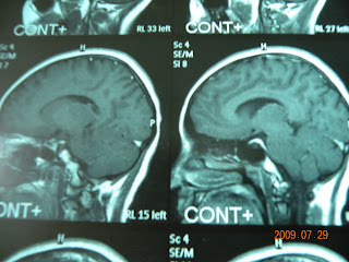
MRI scans


There is confluent area of signal abnormality in left parietal region and basal ganlia. a few small T2 hyperintensities noted on right sided white matter . Post contrast there is patchy enchancement in the centre of this area of signal abnormality measuring around 20*15mm.


Further small enchancing focus noted at the left frontal convexity anteriorly. A third lesion seen on medial aspect of of the base of temporal lobes. Mass effect on ipsilateral ventricular system . no midline shift .

C2-C3 fusion


Hanging out with friends


Saturday, July 18, 2009

Spinal Dysraphism


Multiple cases of spinal dysraphism Meningocele, myelomeningocele and lipomyelomeningocele
operated today. All of them were infants with limb weakness and the congenital defect at birth.
One of them presented with tethered cord syndrome.

Friday, July 17, 2009

TRIGEMINAL NEURALGIA in KEMU


Think of excruciating pain paroxysmal attack very severe intense sharp shooting pain along the branches of Trigeminal divisions in face. Carbamazepine for months and years and still no relief.

Microvascular decompression is the treatment of choice. 100 percent result in our hospital.

Till date we have done and followed 10 patients. All in right side and mostly involving ophthalmic and maxillary divisons. In 9 of them Superior cerebellar artery was the offending vessel and in one case it was unnamed vessel . Result was no neuralgic pain in patients immediate post op. 3 developed hearing impairment but all recovered within few months.

So MVD is such a remarkable operation with 100 % cure rate in our setup.

All the patients underwent surgery under GA in lateral decubitus position neck and head slightly flexed and rotated at 15 degrees towards the contralateral position . The approach was infratentorial supracerebellar through a retromastoid keyhole craniectomy . The arachnoid covering the dorsolateral aspect of the cerebral peduncle was first incised from soperior petrsosal venous trunk in a medial direction parallel to tentorium incisura taking care not to insult the trochlear nerve then laterally and inferiorly to VII-VIII nerve complex . The superor petrosal vein and tributaries dissected free from the surrounding arachnoid membranes and preserved as much as possible. In all patients trunk and 3 main afferents mesencephalic , cerebellar and pontine veins respected . The aim was to preserve the venous return from the cerebellopontine angle, to avoid stretching of VII-VIII nerve complex . The entire trigeminal root was dissected free by dividing the arachnoid filaments and examined from the porus of meckels cave to root entry zone (REZ), so that all neurovascular conflicts could be identified .
Prolene mesh was kept between the Trigeminal nerve root and the offending vessel .