


Cerebelar tonsil retracted
Brain stem visualised nicely
PICA is seen in the operating field


midline incision from inion to level of C2
Occiput and 1st dorsal arch and 2nd cervical spine visualised


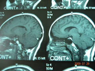


This is a case of 16 yrs old female presenting with headache , dizziness for 1 yr .
Case was operated and a firm mass was visualised in fourth ventricle reaching up t0 mid brain .
Suboccipital craniectomy was done and 1st dorsal arch also removed Y shaped incision over dura . Cerebellar tonsil retracted and vermis exposed and retracted tumor mass was visualised and total resection of tumor from ventricular bed along with its extension achieved .

No comments:
Post a Comment